


21/12/10
Microscope scanner comparison for hematologists and hematopathologists
Models for routine diagnostics tested by Martin Weihrauch, M. D.
The desire to digitally microscope blood and bone marrow smears was the initial impetus for my intensive involvement with digital microscopy almost ten years ago. This is how the company Smart In Media, which I founded as a hematooncologist, became a specialist in this field. In the meantime, we have become the market leader in the field of digital pathology with our PathoZoom® products. However, digital solutions have still not found their way into the routine diagnostics of us hematologists.
This is mainly due to the lack of fast scanners for routine diagnostics in hematology. While our colleagues in pathology have long been able to enjoy good and fast devices with a resolution of 40x and scan times of about 1 minute per slide diagnostics, we hematologists still need to be patient to see our beloved slides digitally.
Even though the skilled pathologist will now probably roll his eyes at the claims of hematologists: Yes, we want a scan that makes our cytomorphology appear digital as if we were viewing the slides with an oil immersion objective at 60x, 80x or 100x in our microscope.
Manual scanning at the microscope in fantastic quality
This is already technically possible today, e.g. with our self-developed manual scanning solution PathoZoom® Scan in fantastic quality. Or also with the Olympus VS120 scanner. However, both options have the limitation that scanning dozens of slides per day would take forever. The Olympus VS120 needs about 30 minutes or longer for a scan of about 1×2 cm. A whole slide would take hours if necessary.
To illustrate how beautiful such a scan with our low-cost PathoZoom® Scan or with the Olympus VS120 can look, I have included cytomorphological slides for you to microscope.
Regarding manual scanning with PathoZoom® Scan, it is important to mention: for scanning a 40x oil with high aperture is sufficient, because the PathoZoom® camera has a higher resolution than the human retina, therefore you get almost the same result as if you were scanning at 100x oil.
Scan with PathoZoom® Scan in oil, 40x Zeiss objective – diagnosis “Malaria tropica”:
Scan with PathoZoom® Scan in oil, 40x Zeiss objective – diagnosis “Normal bone marrow”:
Scan with an Olympus VS120 in oil, 63x objective, diagnosis MDS RAEB-2:
Thus, a scan time of more than 3-5 minutes for an entire blood or bone marrow smear is unacceptable for routine diagnostics. For the spoiled hematologist‘s eyes, there will hopefully be a solution in the near future, a scanner from the American manufacturer Bionovation, which Smart In Media will distribute in Germany.
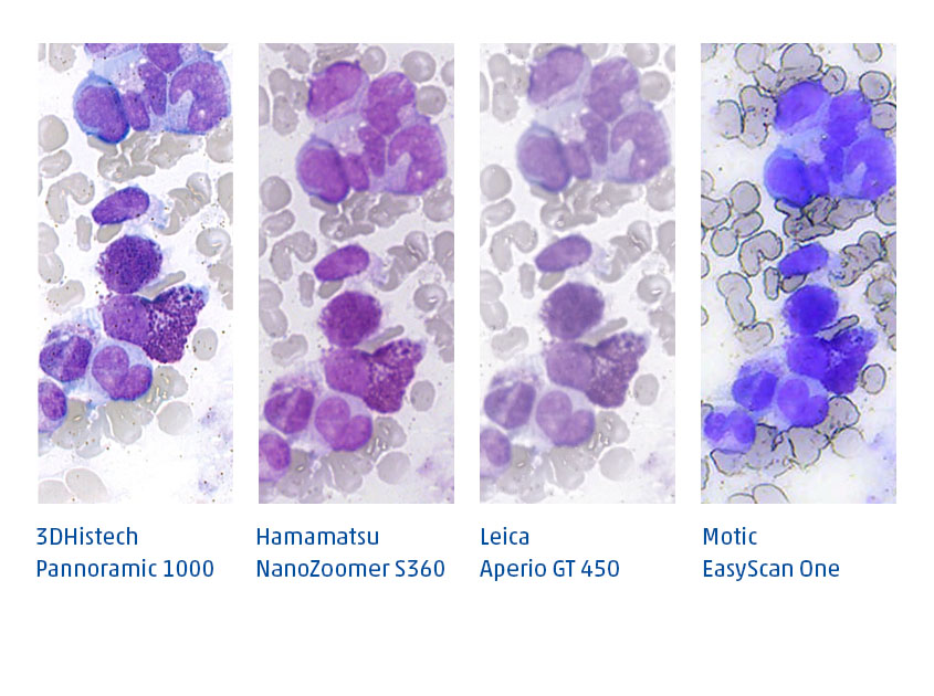
Comparison of three scanners for routine diagnostics
However, there are applications where the quality of the pathology scanners might be sufficient, e.g. for analysis using artificial intelligence.
There are currently (as of 7th Dec 2021) 3 scanners that play a role in routine pathology diagnostics because:
a) they have very good image quality and
b) they have a sufficiently high scanning speed.
These are:
- Hamamatsu – NanoZoomer S360
- Leica – Aperio GT450
- 3DHistech – Pannoramic 1000
Our Smart In Media team has loaded these three scanners with identical specimens (source: Prof. Kreuzer, University Hospital Cologne) and performed scans. In addition, we tested the slower but less expensive device Motic EasyScan One.
All the scanners have advantages and disadvantages in handling. In terms of speed and price, the Hamamatsu S360 is currently ahead. All 3 scanners can be purchased at competitive prices from Smart In Media.
Excellent software from the market leader
In order to be able to view digital slides in routine diagnostics quickly and efficiently, excellent software is of course required, which we offer with PathoZoom® Digital Lab and which is the leading image management system. For more information, please visit our website or contact us.
Regarding the image quality when scanning these slides, I deliberately do not want to influence you. Please form your own opinion!

Below you will find four links, each leading to an order in the PathoZoom® SlideCloud. Each contains identical scans of one scanner.
For those who just want to get a quick overview, I have placed an identical location of a bone marrow preparation from all four scanners side by side.
I wish you joyful microscoping!
Yours
Martin Weihrauch

Up to date via newsletter
Current trends in digital pathology, product news, expert and user meetings, tips and tool recommendations, job offer: Stay up to date with our newsletter. Register here:
