Everyone is talking about digital pathology. However, many questions remain unanswered, especially among newcomers. In our interactive DIGITALIZATION PRIMER, Martin Weihrauch, M. D., shows how pathologists can make their institute fit for the future with digital diagnostics.

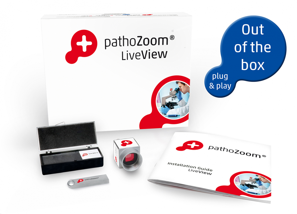
The Digitization Primer is only available from us!

We would also be happy to send you a printed copy ($6/€5 plus postage); to do so, please send an e-mail with your address to Corina Turner, e-mail c.turner@smartinmedia.com.

Digital Pathology – Questions & Answers

Pathologe am klassischen Mikroskop

Digital Pathology saves time, reduces costs, and increases quality in pathology in various situations.

Even with just a small investment, physicians and medical assistants at frozen section sites or even off-site locations can exchange information about a case digitally – thus in real-time – with specialist colleagues.

Microscopes can be converted to digital diagnostics by using microscope digitizers and a corresponding image management system. In this way, pathologists save time, as digital microscopy can be faster than using a standard microscope nowadays.

You already have everything on-site at your institute for this: a microscope and a computer with internet access are certainly available at your frozen sectioning station.

For the first steps into digital diagnostics, we equip your microscope with a high-speed camera and your computer with our PathoZoom® Scan & PathoZoom® LiveView software. This robust system is immediately ready for use: You can scan on your own microscope.

Physicians and MTAs at the frozen section station can connect to you immediately with this starter package, without having to install any software since everything runs via the browser on your PC/laptop, tablet or smartphone. With any of these devices, you see exactly what your co-workers see on the microscope in the lab at your workstation, at home or on the road!

Produktverpackung Scan und LiveView

With this package, you can also digitally scan slides manually on your microscope – at exactly the same resolution as an expensive scanner. You are able to share the digitized slides with colleagues within seconds via PathoZoom® SlideCloud, an image storage space reserved just for you – simply with a link via email or messenger.
You get the whole thing for less than $17,000!

By the way, PathoZoom® Scan & PathoZoom®LiveView are installed on hundreds of microscopes worldwide. This improves communication among pathologists enormously, including
many colleagues who are diagnosing at this moment!

Pathologists that successfully digitize are changing their processes step by step with our guidance. We recommend buying or renting the minimum equipment to start.

This allows you to test digital diagnostics at your own pace and avoid high initial investments. In addition, the field of Digital Pathology is currently in the fast lane: Equipment and systems that you buy today may be obsolete tomorrow.

For the beginning, just one lab technician and one physician should start working with the system, not the entire staff right away. That way, you can digitize the workflow in your institute step by step in a deliberate way and bring your experience to the next stage of the transition.

You already have …

  • a Laboratory Information System (LIS)

You need …

  • a diagnostic platform with a viewer, e.g. PathoZoom® Digital Lab
  • one/several high-performance scanner(s)
  • a printer with which you can directly print your slides or labels with a machine-readable code (DataMatrix code). For this purpose, you can use the easy-to-use software PathoZoom®LabelPrinter plus a printer
  • a computer and storage space for the slides

Possible uses of the PathoZoom Digital Lab diagnostics platform

The most important thing for digitization is choosing the correct diagnostics platform. It replaces your microscope at least partially and connects your Laboratory Information System (LIS) and your scanner(s).

It is crucial that your diagnostics platform is an OPEN system. Open to all scanners, open to all LIS, open to Artificial Intelligence and above all: open to your IT department.
Ensure that you or your IT retain complete access to the digital slides, the database, and the interfaces.
The interface of your diagnostic platform should be free of charge (not priced!). It must also be able the read the DICOM files – the future open file format.

PathoZoom® Digital Lab fulfills all these requirements. The diagnostic platform is the heart of digitization and connects all critical digitization components of your lab. The open architecture ensures the future viability of your complete digital system.

Because PathoZoom® Digital Lab is:

  • open for all scanner systems and formats
  • open for all LIMS systems
  • open for any data storage (local or in the cloud)
  • open for the integration & training of all AI modules

Ideally, you have two screens: One shows your Laboratory Information System (LIS), and the other shows your PathoZoom® Digital Lab as a digital microscope and diagnostics platform.
PathoZoom® Digital Lab does not have to run on the same computer as your LIS.

You can also use a modern tablet computer as a digital microscope since you do not need to install any software for PathoZoom® Digital Lab. This allows you to microscope on any device with a browser and the Internet.

If you open a case in the LIS, the same case opens in PathoZoom® Digital Lab, including the corresponding folder with all associated slides.

See for yourself: Click here or scan the QR code on the right side now with the camera function of your smartphone.

  • With your smartphone, at the top right, you will see three horizontal lines. If you click on these, you will find the
    microscope folder. You can microscope with your finger, e.g., zoom into the slide with your thumb and index finger,
    use the measuring tool, annotate, etc.
  • If you would like to test the PathoZoom® Digital Lab on your large computer screen, forward the link to your
    desktop PC where you can work with the mouse as usual.

How is this working for you?
Our team, physicians and developers, are looking forward to your feedback to info@smartinmedia.com.

The purchase of one or more scanners comes with high costs. In addition to the classic purchase, there are also rental and leasing models.

If you have an image management system that can handle ALL scanner formats, all scanner brands are available to you.
You can combine scanners from different companies in one system.

As of 2022, there are three companies whose scanners we recommended for routine use: Hamamatsu, Leica and 3DHistech. You should test each of these scanners before making a purchase. All are very good in quality but differ in their workflow, handling, and price – and every pathologist has different requirements.

Smart In Media has a lot of experience with scanners and numerous customers with different devices, and will gladly give you an on-site inspection.

Please call us at +49 221 999943-00

You can find out more about scanners in our scanner test here.

We are the solution partner for pathologists worldwide – not just an ordinary software company. We take full responsibility for ensuring that all our components work together at your site.
In addition: Our PathoZoom® Digital Lab server, installed in your institute, monitors all the slides digitized by the scanner and automatically groups them into cases.

Several physicians work in and for our company. Together we live the philosophy “by physicians for physicians.” This results in our simple workflow and many helpful details, including useful tools like the PathoZoom® Spacemouse and the PathoZoom® LabelPrinter.

In addition, we have interfaces to all common LIS providers, so that cases you open in your LIS also immediately appear in PathoZoom® Digital Lab.

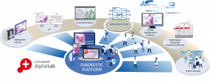
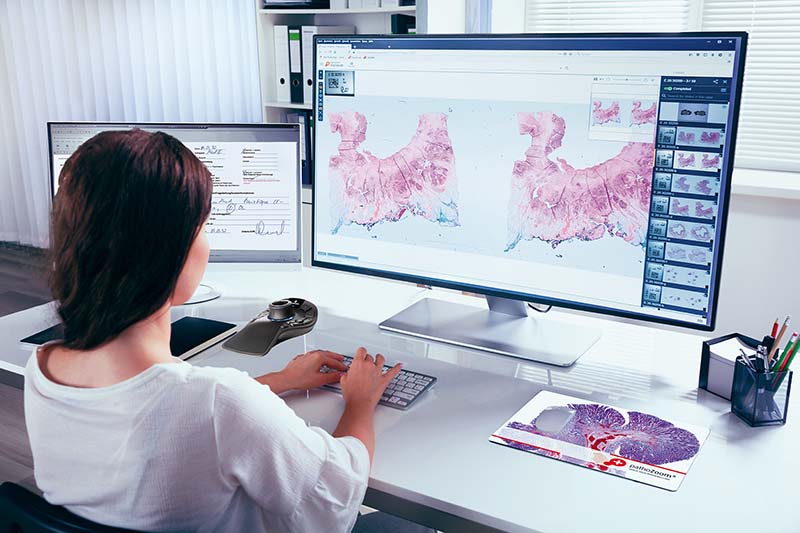
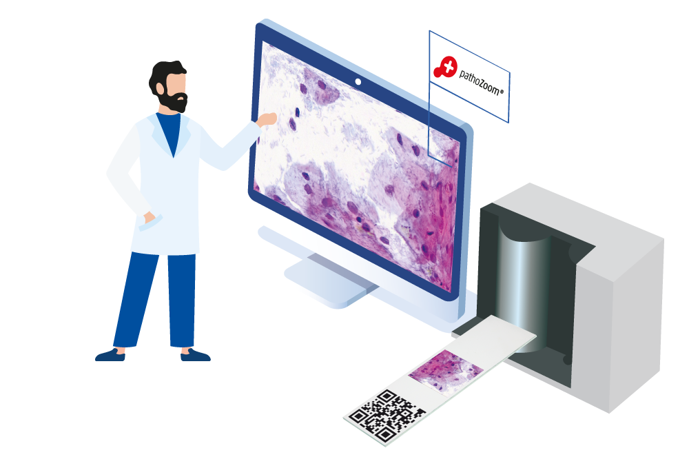
When comparing the classic to the digital workflow, these steps change:

  • The slides are provided with a 2D Barcode for digitization, typically with a DataMatrix Barcode so that PathoZoom® Digital Lab can later assign the slides to the cases automatically. The slides are provided with tissue and stained as usual.
  • The slides no longer have to be sorted, they can be scanned in any order.
  • There is no longer any need to stack folders with slides to individual pathologists; your lab technicians can quickly assign cases in your Digital Lab.
  • While the first cases are still being scanned, the pathologist already begins their reporting.

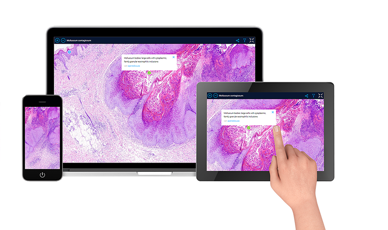
First of all, you can already start reporting parallel to the scanning process. In addition, you save time with the following features of Digital Pathology:

  • Digital microscopy on-screen is faster and more precise than on the microscope (changing slides, jumping between cases, …).
  • You save time through faster retrieval of structures.
  • There is no need to search for/mix up slides and cases, saving you time and annoyances.
  • Case acceptance by assistants is accelerated many times over, as they can already mark case-critical regions (e.g., Helicobacter pylori, invasion front, etc.).
  • Communication with colleagues is accelerated through shared digital microscopy.
  • You can easily involve pathologists at remote sites.
  • Retrieval of old cases – with long-term archiving – is possible instantly.

Digital diagnostics offers many other improvements:

  • Barcodes increase the security regarding the correct assignment of slides. The pathologist no longer needs to “read” the slides.
  • Cases can also quickly be shared with external pathologists (reference pathologists and others) by mouse click.
  • You can quickly present cases digitally at the tumor board.
  • Macroscopy and microscopy can conveniently be combined.
  • Working at the screen is more comfortable than at the microscope due to sitting upright and eliminating focusing back and forth between the request form, slide folder, and microscope.

Currently (as of 2022), more and more Artificial Intelligence (AI) developments are entering the market. One challenge is that in the future, as a pathologist, you will not need just one Artificial Intelligence package, but many. There will be one for every DX, such as finding metastases in lymph nodes in colon carcinoma, analyzing PD-L1 status, and much more.

You can compare future AI to the antibodies for immunohistochemistry that you use today. There is an advantage here, too, if you can freely choose between different vendors later on.

With each AI, you would need a separate server installation from this special vendor. We have solved this with PathoZoom® Digital Lab and the integrated “AI App Store” just for Artificial Intelligence. With it you can freely and without any installation effort book or switch different AI applications.

  • Scientific publications and studies on our systems can be found here
  • Use cases of our digital solutions from universities, clinics and institutes are compiled here.
  • Book your visit to a laboratory of one of our reference customers via our contact form or by phone +49 2221 999943-00
  • Exchange information about digital diagnostics with colleagues worldwide at our interactive expert meetings – here are the dates and topics of our next events
  • Book your preferred date for a demo or consultation for the best start via our contact form or by phone +49 221 999943-00

Call us for a free expert consultation. The most helpful way to get started with Digital Pathology is to talk directly with our doctors and specialists.

If you are interested, we would like to invite you to visit one of our reference customers, where you can experience digitized workflows live and exchange ideas with colleagues on site.

Call us, send us an email, or visit us:

Smart In Media AG
Gleueler Straße 249, 50935 Cologne/Germany
Tel. 0221 999943-00
info@smartinmedia.com

USA Office
14460 Falls of Neuse Road, Suite 149262, Raleigh, NC, 27614
Don van Dyke, Vice President, GM
Phone: +1 (919) 345-2838
d.vandyke@smartinmedia.com

Of course, we are also personally available to answer all your digitization questions via our contact form or by phone +49 221 999943-00.