
2 modes
for every situation of your workday
5 minutes
of installation, and you’re ready to go
less than 60
seconds to scan a whole specimen

AI-Powered Image Analysis – When You Need It
- Integrate AI modules seamlessly into your workflow for tasks like cell counting and biomarker analysis
- View results instantly, directly within your image
- Supports established markers such as Ki-67, ER, PR, PD-L1, and more
- Automatically store AI results alongside your case data for full traceability
Plug & Play – Ready in Minutes
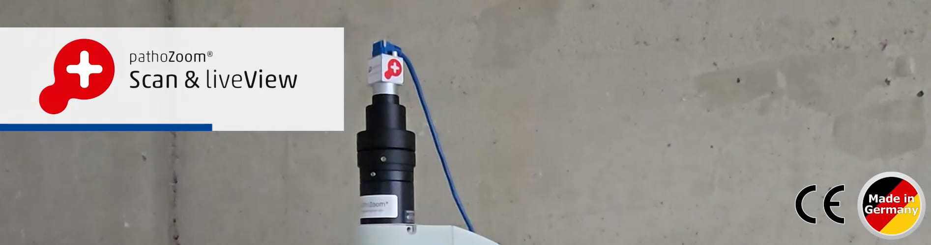
- Compatible with any standard microscope via C-mount
- Set up in minutes – no IT expertise required
- All-in-one package: CE-certified software, high-resolution camera, calibration slide, and remote support
- Upgrade your existing microscope into a digital solution – quickly and effortlessly
Popular Use Cases
- Diagnostics: Frozen sections, second opinions, tumor boards
- Education & Training: Interactive digital microscopy for students and lab professionals
- Research & Pharma: Image analysis and efficient data sharing in clinical studies
- Telepathology: Collaborate remotely with partner labs and international experts
- Documentation: Create and manage digital slides for archives, reports, and presentations
What’s in the Box?
- PathoZoom® USB 3.0 microscope camera & cable
- CE-certified PathoZoom® Scan & LiveView software
- Calibration slide for precise measurements
- Remote installation and onboarding support
- Compatible with all Windows PCs and common microscopes with C-mount adapter
PathoZoom® Scan & LiveView is IVDR-compliant and features CE-certified measurement tools for reliable diagnostic use.

“Digital pathology brings many advantages to physicians and patients. The most important thing is to take the leap and get started”