
𝗔𝗻𝗱 … 𝗔𝗰𝘁𝗶𝗼𝗻: 𝗗𝗶𝗴𝗶𝘁𝗮𝗹 𝗗𝗲𝗿𝗺𝗮𝘁𝗼𝗵𝗶𝘀𝘁𝗼𝗹𝗼𝗴𝘆 𝘄𝗮𝘀 𝗻𝗼𝘁 𝗷𝘂𝘀𝘁 𝗱𝗶𝘀𝗰𝘂𝘀𝘀𝗲𝗱 𝗮𝘁 𝗔𝗗𝗛.
It was demonstrated and put into practice.
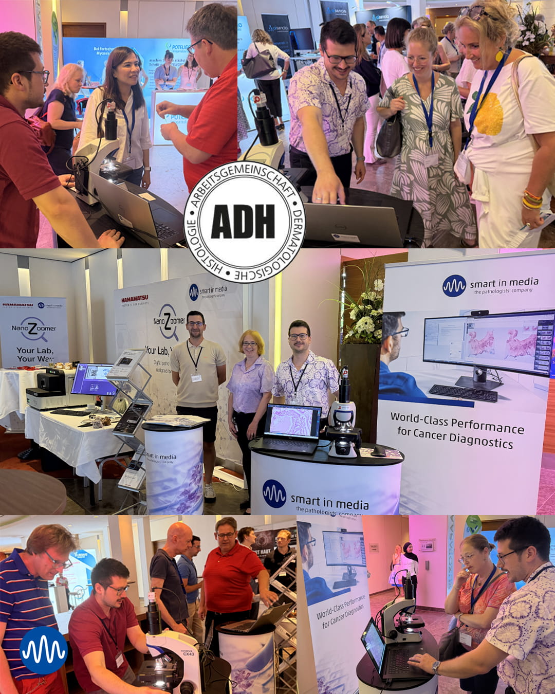
At the Annual Meeting of the Working Group for Dermatological Histology (ADH) in Münster, Corina Turner, Daniel Becker, and Ralph Gloser brought digital diagnostics to life on site. They demonstrated live how broadly digital dermatohistology can be approached today: from fast digitization directly at the microscope, to LiveView scenarios for consultations, exchange, and teaching, all the way to a fully digital workplace with scanner, image management system, diagnostic platform, and openness for AI.
This practical perspective made our joint booth with Hamamatsu Photonics Deutschland GmbH a busy meeting point throughout both days. The many conversations clearly showed how strongly dermatohistology is currently moving toward digital workflows — not everywhere at the same pace, but with increasingly concrete plans.
Some institutes and practices are starting with specific applications directly at the microscope. Others are already planning end-to-end workflows — from scanner and IMS to AI-supported diagnostics and reporting processes.
This is not just about digital images. It is about diagnostic work that can become more structured, better connected, and more efficient. In suitable workflows, digital pathology can reduce diagnostic reporting times by 12–30% — provided that scanner, image management system, diagnostic platform, and processes work together in a meaningful way.
And that was exactly the focus: demonstrating, trying out, and putting things into perspective.
Up to date via newsletter
Current trends in digital pathology, product news, expert and user meetings, tips and tool recommendations, job offer: Stay up to date with our newsletter. Register here: